I was fortunate enough to work with Dr. Terri A. Williams at my alma mater, Trinity College in Hartford Connecticut studying the Evolutionary Developmental Biology of arthropod segmentation.
Like us vetebrates, arthropods are segmented (think of a centipede!), and like vertebrates, many arthropods build segments during developmentment sequentially. In my previous work. I've investigated (Nakomoto et al., 2015) the process of segmentation and elongation in a short germ insect the Red Flour Beetle (Tribolium castaneum). Like most sequentially segmented organisms, Tribolium generates new segments during development from the posterior, undifferentiated zone called the growth zone (which is the region posterior to the final engrailed stripe).Previous research has implicated Wnts, even skipped/odd skipped, and caudal as being involved in a network that regulates formation of new segments; however, it is unclear how universal this gene regulatory network is within growth zones among arthropods.
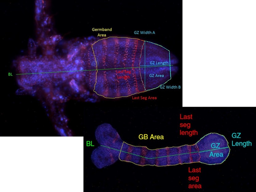
Taking a comparative approach, I began to study a close relative to Artemia, or "Sea Monkeys", a branchiopod crustacean, Thamnocephalus platyurus, to see how similar the process of segment formation is within arthropods. Starting with simple measurements between embryos at different stages of development, we were able to characterize the growth zone and how it changes over the course of segmentation in Thamnocephalus (top) and Tribolium (bottom)
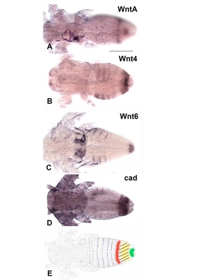
Collaborators and I created a trancriptome and documented the Wnt genes in the growth zone (and in the rest of the body) of Thamnocephalus, suggesting different Wnts may be at play. Unlike Tribolium which undergoes segmentation within the egg, Thamnocephalus hatch with three segments (of the 19 they have as adults) and are free swimming and feeding as they undergo both segmentation and morphogenesis. Dr. Williams is continuing to explore how differences in phylogeny and life history may affect growth zone characteristics among arthropods.
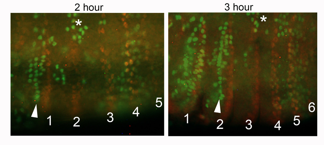
Dr. Williams and I are currently preparing another manuscript to document growth zone gene expression and to determine the relative contribution of cell movements and mitosis to segmentation in Thamnocephalus. The image below depicts the changes in mitotic cells (as measured by cells cycling through S-phase- green staining of EdU) over the course of ~1 hour when a new segment is formed. The posterior of the segment is highlighted in red and numbered, "*" denotes the neuroectoderm, note the posterior progression, and the arrowhead highlights a pattern of mitosis where the apical ridge of cells in the segment coordinate their divisions in order to elongate the segment into what will be the adult paddle-like limbs. Anterior is to the left and this is the ventral surface of the body.
Nakamoto, et al., 2015. Changing cell behaviors during beetle embryogenesis correlates with slowing of segmentation. Nature Communications 6:6635 doi: 10.1038/ncomms7635
Constantinou, et al., 2016. Wnt repertoire and developmental expression patterns in the crustacean Thamnocephalus platyurus. Evolution & Development18: 324-341. doi: 10.1111/ede.12204